Diagnostic Imaging
- About the Observatory
- Team
- Units and benefits
- Diseases and procedures
- Research
- Teaching activity
- Related information
About the Observatory
The Imaging Diagnostic Service (SDI) belongs to the so-called central services of a hospital together with Laboratory, Pathological Anatomy and Pharmacy, which support the other specialties.
Its main activity consists of:
- Obtaining anatomical images using different devices-modalities that use various physical methods, in order to reach a diagnosis or confirm normality (eg: taking radiographs using X-ray devices, ultrasound, computed tomography, magnetic resonance).
- Obtaining, with the support of the different modalities-devices, biological samples for clinical, microbiological or anatomopathological analysis (eg: puncture-aspiration or biopsy of tumor lesions, puncture-aspiration of body fluids).
- Performing therapeutic procedures or to support future treatment, guided by the image (eg: drainage of abscesses, placement of catheters, placement of harpoons or breast markers prior to surgery).
He is also part of several interdisciplinary committees where clinical cases are assessed in order to assess the attitudes to be followed.
The human team of the SDI is made up of: doctors specializing in radiodiagnosis, nurses, senior technicians in diagnostic imaging, nursing assistants, stretcher carriers and administrators, all of them in charge of one or more tasks within the different processes that make up the global activity of the service
-
Update date: 01.01.2024
Head of Service

-

Jordi Catala Forteza
diagnosis by hgh image
Head of Service
-

Maria Del Pilar Lozano Arranz
diagnosis by hsjd image
service coordinator doctor
-

Jose Maria Vila De Miguel
diagnosis by hsjd image
service coordinator doctor
-

Sonia Ruiz Macarrilla
diagnosis by hgh image
service coordinator doctor
-

Javier Miguez Gonzalez
diagnosis by hsjd image
specialist doctor
-

David Esteban De Bonadona Tramon
diagnosis by hsjd image
specialist doctor
-

Javier Milian Garcia
diagnosis by hsjd image
specialist doctor
-

Gracia Valderas Martinez
diagnosis by hgh image
specialist doctor
-

Eva Almazan Mesa
diagnosis by hsjd image
specialist doctor
-

Francesc Calaf Forn
diagnosis by hsjd image
specialist doctor
-
Ingrid Carolina Duran Palacios
diagnosis by hsjd image
specialist doctor
-

Rafael Oliveira Caiafa
diagnosis by hsjd image
specialist doctor
-
Diego Solis Gutierrez
diagnosis by hsjd image
specialist doctor
-

Maria Catalina Montull Ferrer
diagnosis by hsjd image
specialist doctor
-

Vicente Valles Noguero
diagnosis by hsjd image
specialist doctor
In this section appear the professionals of the Integral Health Consortium who have authorized the display of their personal data.
-
Update date: 01.01.2024
They allow the so-called conventional radiology to be carried out, consisting in obtaining radiographs of any part of the body through the emission of X-rays.
Scans distributed by sections of organs and systems:
- Bodysuit: section aimed at the study of the pathology of the thoracic and abdominal cavities and thyroids.
- Chest:
- Chest radiography: continues to play a primary role in the initial study of numerous pathologies of both the respiratory and cardiovascular systems. Required in some patients for preoperative assessment.
- Abdomen:
- Abdominal X-ray: it has a limited role practically in the field of emergencies as the first study in the case of suspected intestinal obstruction or in the study of lithiasis pathology of the urinary tract.
- Chest:
- Musculoskeletal: section aimed at the study of bone, musculotendinous and joint pathology
X-rays of all the bones and joints of the body including scoliograms and telemetry - Vascular-Interventionist: section aimed at carrying out vascular studies and interventionism of medium complexity
The device consists of a mobile table in the 3 axes of space and a mobile X-ray tube in the longitudinal axis, which allows, in addition to conventional radiology, to carry out dynamic radiological studies prior to the introduction of material from contrast either oral or rectal, intravenous, endocavitary or intra-articular.
Tests with oral contrast: esophagogram, esophagogastroduodenal transit, intestinal transit
Rectal contrast tests: opaque enema
Tests with intravenous contrast: intravenous urography, phlebography, arteriography
Tests with endocavitary contrast: hysterosalpingography, cystography, urethrography, fistulography
Tests with intra-articular contrast: arthrographies
It is also possible to check the permeability of structures or catheters by administering contrast through them as in trans-Kher cholangiography or pyelography for nephrostomy.
We also have a multi-function remote control that allows interventional procedures of medium complexity such as percutaneous biliary drainage or the placement of tunneling catheters for dialysis. It has videofluoroscopy that also allows dynamic studies such as the swallowing test.
Scans distributed by sections of organs and systems:
- Bodysuit: section aimed at the study of the pathology of the thoracic and abdominal cavities and thyroids.
- Chest: Thoracic endoscope: aimed at assessing diaphragmatic movement.
- Abdomen: Tests aimed at studying the digestive system:
- Esophagogram: with administration of oral contrast (barite or iodide) for the study of organic and functional pathology of the esophagus.
- Esophagogastroduodenal transit: with administration of oral contrast (barium or iodide) for the study of pathology of the first section of the digestive system.
- Intestinal transit: with the administration of barite oral contrast aimed at the study of the pathology of the small intestine.
- Opaque enema: with rectal administration of contrast (barite or water-soluble iodide) for the study of the pathology of the large intestine.
- Swallowing test: study using videofluoroscopy of swallowing motility with the administration of barite oral contrast preparations.
- Tests aimed at studying the urinary system:
- Urography: with administration of intravenous iodinated contrast to study the urinary tract.
- Cystography and cystourethrography: with endocavitary iodinated contrast administration for the study of the urethra and urinary bladder.
- Trans-Kher cholangiography: evaluation of the biliary tract with the injection of iodinated contrast through a Kher tube.
- Pyelography for nephrostomy: evaluation of the urinary tract with the injection of iodinated contrast through a nephrostomy catheter.
- Musculoskeletal: section aimed at the study of bone, musculotendinous and joint pathology
- Arthrographies: previous injection of intra-articular iodinated contrast
- Pathology of women: section aimed at the study of the specific pathology of gynecological and mammary women
- Hysterosalpingography: Test aimed at evaluating the uterine cavity and fallopian tubes prior to the administration of iodinated contrast to the same endometrial cavity.
- Vascular-Interventionist: section aimed at carrying out vascular studies and interventionism of medium complexity
- Arteriography: arterial study of extremities
- Phlebography: venous study of extremities
- Nephrostomy: placement of percutaneous drainage in the urinary tract
- Biliary drainage: placement of percutaneous drainage in the biliary tract
- Tunneling of vascular accesses for dialysis
It uses X-rays. Mammograms are obtained specifically and allows interventional procedures to be carried out using the stereotaxic system such as biopsies or placement of harpoons prior to surgical intervention, and checking of surgical parts. Galactography is also carried out, which consists of the study of the pathology of the galactophorous ducts after the introduction of iodinated contrast inside them.
Scans distributed by sections of organs and systems:
- Pathology of women: section aimed at the study of the specific pathology of gynecological and mammary women
- Mammograms: aimed specifically at the study of breast pathology.
- Galactography: study aimed at studying the pathology of the galactophorous ducts prior to the introduction of iodinated contrast inside them.
- The stereotaxic device integrated in the mammograph itself allows interventional procedures to be carried out:
- Stereotaxically guided coarse needle biopsy
- Placement of harpoons guided by stereotaxy or by coordinates.
- Checking of surgical parts
- Mammograms: aimed specifically at the study of breast pathology.
It obtains images using ultrasound, which have no harmful effects, of any part of the body with limitation of bony structures and some degree of limitation with aerated digestive structures or very deep localization. The Doppler mode allows the dynamic study of flows in the vascular structures. It is used very frequently as a support for interventional procedures especially on superficial structures (fine needle punctures (FNA), coarse needle biopsies (BAG) and placement of catheters).
Scans distributed by sections of organs and systems:
- Bodysuit: section aimed at the study of the pathology of the thoracic and abdominal cavities and thyroids.
- Chest:
- Thoracic ultrasound: aimed mainly at the study of pleural effusion
- Ultrasound is also used as a guide for performing various interventional procedures:
- Thoracocentesis: both diagnostic and evacuation where a drainage catheter is often left in place in the latter.
- Lung/pleural biopsy: of peripheral lung tumors or pleural tumors.
- Abdomen:
- Abdominal ultrasound: aimed at the study of most of the intra-abdominal organs and for the detection of free fluid, with a limitation towards the digestive system.
- Reno-vesical and reno-vesico-prostatic ultrasound: aimed at the study of renal, bladder (mainly tumor and assessment of emptying) and prostatic (mainly assessment of hyperplasia) pathology. Note that abdominal ultrasonography is not an optimal method for assessing prostatic tumor pathology.
- Renal ultrasound: evaluation of the kidney.
- Thyroid and neck ultrasound: the body section also undertakes studies aimed at ruling out thyroid pathology and cervical structures.
- Ultrasound is also used as a guide for the performance of various interventional procedures, especially on organs and structures of superficial localization:
- Puncture-aspiration with a fine needle.
- Biopsies of any solid organ including the prostate using a transrectal transducer.
- Placement of drains for intra-abdominal collections or organs:
- Cholecystostomy: specific drainage of the gallbladder
- Nephrostomy: specific drainage of the urinary tract
- Chest:
- Musculoskeletal: section aimed at the study of bone, musculotendinous and joint pathology
- Ultrasounds aimed mainly at the study of musculotendinous lesions, of soft parts and periarticular pathology. The most requested is the shoulder ultrasound.
- Ultrasound is also used as a guide for various interventional procedures, puncture-aspiration, biopsies and extraction of foreign bodies.
- Neuroradiology: section aimed at the study of the pathology of the central nervous system, head and neck
- Ultrasounds: PAAF-Biopsy of cervical lesions/adenopathy
- Pathology of women: section aimed at the study of the specific pathology of gynecological and mammary women
- Breast ultrasound: aimed at the study of focal lesions
- Axillary ultrasound: aimed at assessing the axillary nodes to stage breast cancer.
- Ultrasound is also used as a guide for performing various interventional procedures:
- Ultrasound-guided puncture-aspiration
- Ultrasound-guided coarse needle biopsy
- Placement of breast harpoons and markers guided by ultrasound prior to surgery.
- Evacuation puncture of mammary collections and placement of drainage catheters.
- Vascular-Interventionist: section aimed at carrying out vascular studies and interventionism of medium complexity
- TSA Doppler ultrasound: study of atheromatous pathology of the supraaortic trunks
- Doppler ultrasound of renal arteries: for assessment in the context of arterial hypertension
- Doppler ultrasound of limb arteries
- Venous Doppler ultrasound of the extremities: for assessment of thrombotic pathology and venous insufficiency
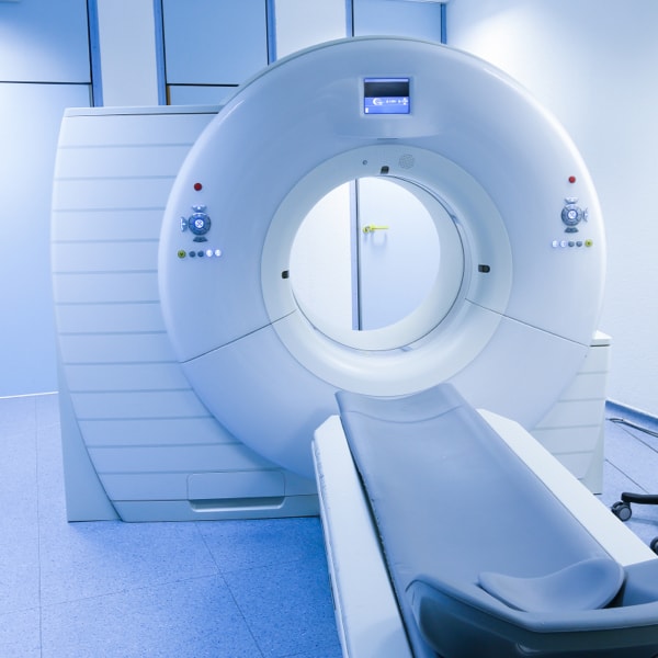
It obtains images of any part of the body using an x-ray tube that rotates axially toward one or more rows of detectors while moving the table on which the patient lies. The number of detectors will determine the capabilities of CT in terms of image acquisition speed characteristics and their thicknesses. The administration of intravenous iodinated contrast increases the sensitivity and specificity of tests towards certain types of pathology. Likewise, some studies may require the administration of oral or rectal contrast or previous cleaning of the digestive tract. It is used with some frequency as an adjunct to interventional procedures (fine needle aspiration (FNA), coarse needle biopsies (BAG) and catheter placement).
Scans distributed by sections of organs and systems:
- Body: section aimed at the study of the pathology of the thoracic and abdominal cavities and thyroids.
- Chest:
- Chest CT: mainly aimed at the study of airway pathology, lung nodule study and tumor staging.
- High-resolution chest CT: mainly aimed at studying the pathology of the pulmonary interstitium.
- Angio-CT of pulmonary arteries: to rule out pulmonary thromboembolism.
- Angio-CT of aorta: for study of thoracic aorta.
- Cardio-CT of coronary arteries and "calcium score": specific study of the coronary arterial tree.
- Lung biopsy: CT is used as a guide for performing lung biopsies.
- Abdomen:
- Abdominal CT: mainly indicated for the staging of tumors, characterization of focal lesions of any intra-abdominal organ and evaluation of the retroperitoneal space.
- Uro-CT: aimed at the study of the pathology of the lithiasis or tumoral urinary tract.
- Colono-CT/Virtual Colonoscopy: aimed at the study of colonic tumor pathology mainly in those cases with contraindications to colonoscopy or when it is incomplete. Administration of oral contrast is required to mark the stool.
- CT is also used as a guide for various interventional procedures, punctures, biopsies and placement of drainage catheters.
- MR abdomen: mainly for the study of focal lesions of any intra-abdominal organ. Quantification of iron in the liver in suspected or studied hemochromatosis.
- Pelvic MRI: for the study of focal lesions in female pelvic organs, staging of rectal neoplasia and study of recto-anal inflammatory pathology (fistulas).
- Cholangio-MR: aimed at the study of the biliary tract.
- Entero-MR: aimed at the study of intestinal inflammatory pathology.
- Uro-MR: assessment of urinary tract pathology.
- MR-prostate: specific evaluation of the prostate gland mainly for the detection of tumors.
- Defecography-MR: specific assessment of pelvic floor motility
- Committees: hepato-biliary, carcinomatosis, colorectal cancer, digestive cancer, prostate cancer, kidney-urinary tract cancer
- Chest:
- Musculoskeletal: section aimed at the study of bone, musculotendinous and joint pathology
CT of any bone and joint of the body mainly aimed at the study of bone (fractures and tumors) and joint pathology. Limitation in the study of meniscal, ligamentous and musculotendinous structures.- Dental CT: aimed at the study of dental parts.
- CT is also used as a guide for performing various interventional procedures, puncture-aspiration and biopsies.
- Neuroradiology: section aimed at the study of the pathology of the central nervous system, head and neck
- CT of the head, CT of the neck, CT of the paranasal sinuses, CT of the facial mass, CT of the ears, CT of the orbit, CT of the rock, CT perfusion.
- Intracranial CT angiography, supraaortic trunks CT angiography
- Pathology of women: section aimed at the study of the specific pathology of gynecological and mammary women
- Abdominal-pelvic CT: mainly for staging gynecological and breast tumors
- Pulmonary CT: mainly for staging gynecological and breast tumors
- Vascular-Interventionist: section aimed at carrying out vascular studies and interventionism of medium complexity
- Angio-CT of aorta: specific study of the aorta artery
- Angio-CT of a. mesenteric: specific study of the superior mesenteric artery.
- Angio-CT of a. renal: specific study of renal arteries
- Angio-CT of the lower extremities: of the arteries of the legs
It obtains images of any part of the body with some degree of limitation regarding highly airy structures or with the presence of gas. Obtaining images is based on the different behavior of body tissues under a magnetic field when radio frequency waves are emitted. The administration of paramagnetic contrast with Gadolinium intravenously increases the sensitivity and specificity of the tests towards certain types of pathology. Likewise, some studies may require the administration of compounds orally
Scans distributed by sections of organs and systems:
- Bodysuit: section aimed at the study of the pathology of the thoracic and abdominal cavities and thyroids.
- Chest:
- Cardio-MR: specific study of cardiac pathology. There are several types of tests, some of which will require the administration of drugs or paramagnetic contrast. These tests are performed and assessed together with a cardiologist.
- Types of tests: Functional Cardio-MR, Functional Cardio-MR with study of myocardial viability and/or myocardial perfusion, Cardio-MR for flow study
- Thoracic MRI: MRI has a limited role in the study of thoracic pathology, mainly directed at the study of mediastinal pathology.
- Angi-MR of pulmonary veins: specific study of pulmonary veins
- Committees: lung cancer, interstitial diseases.
- Chest:
- Musculoskeletal: section aimed at the study of bone, musculotendinous and joint pathology
MRI of any muscle group, bones and joints, with good resolution for the study of meniscal and ligamentous structures. The most frequent are:- Knee MRI, Shoulder MRI, Spine MRI, Hip MRI, Ankle MRI, Wrist MRI, Sacroiliac MRI, Elbow MRI.
- Arthro-MR: previous intra-articular injection of paramagnetic contrast (Gadolinium). Mainly wrist, shoulder, knee and hip.
- Brachial plexus MRI: specific study of the brachial plexus nerve roots.
- Neuroradiology: section aimed at the study of the pathology of the central nervous system, head and neck
- Cranial MR, Cranial MR with Spectroscopy, Orbital MR, Spinal MR (for spinal cord assessment), Pituitary MR, Internal auditory canal MR, Penial MR, Cerebrospinal fluid flow MR (CSF dynamics), MR of neck, MR of larynx, MR of oropharynx, MR of parotid, MR of paranasal sinuses.
- Intracranial MR angiography, supraaortic trunk MR angiography
- Pathology of women: section aimed at the study of the specific pathology of gynecological and mammary women
- Breast MRI: specific study of breast pathology, mainly tumor detection, characterization and staging and assessment of prostheses.
- MR pelvis: study of gynecological tumors. Study of the pelvic floor.
- Vascular-Interventionist: section aimed at carrying out vascular studies and interventionism of medium complexity
- Angio-MR of the aorta: specific study of the aorta artery
- Angio-MR of a. renal: specific study of renal arteries
- Angio-MR of the lower and upper extremities: of the arteries of the legs and arms
- MR angiography of visceral arteries: study of abdominal arteries
- Venous MR angiography: specific study of veins
- 3 Conventional radiology devices
- 1 Mammograph
- 1 Multi-function remote control
- 3 sonographers
- 3 CTs (one of 64 and two of 128 detector crowns respectively)
- 1 RM of 1,5 Teslas
Likewise, HMB and HGH have portable devices and surgical arcs for performing conventional radiology in the ICU, for admitted patients who cannot be moved to the SDI and support in the operating room.
The two acute hospitals (HMB and HGH) carry out ambulatory activity in the different modalities from Monday to Friday from 8 a.m. to 22 p.m., activity towards scheduled inpatients and on demand, and urgent activity towards inpatients and emergency rooms 24 hours a day day.
After-hours urgent CTs are reported via teleradiology.
Apart from the CSI centers themselves (HMB, HGH, Hospital Sociosanitari de l'Hospitalet, ABS Ronda Torrassa and ABS Collblanc), coverage is provided on demand at the CAE (Specialized Care Center) in Ronda Torrassa, Cornellà de Llobregat and of Sant Feliu de Llobregat. HADO (Home Hospitalization) and SEVAD (Dependency Assessment Service) are also supported.
The breast cancer screening program of the ICO of the area is also supported with the performance of around 4.000 mammograms per year.
- Urology Committees (Prostate-Kidney/Urinary Tract)
- Otorhinolaryngology Committee
- Lung Committee
- Hepatobiliary Committee
- Carcinomatosis Committee
- Colorectal Committee
- Digestive pathology committee
- Breast pathology committee
- Gynecology Committee
- Mortality Commission
- Commission for the improvement of clinical practice-Guidelines Commission
-
Update date: 01.01.2024
Diseases and procedures
-
It was good that both prescribing doctors and patients knew information about certain aspects related to the tests carried out at the SDI. The demand for these has been growing considerably in recent years, and considering that resources are limited, it is necessary to adjust the demand only when it is really justified. Each test has indications, and what is good for one patient or for the study of a certain pathology is not always good for another. Ultimately, it is the responsibility of the radiodiagnostic specialist to approve the requested tests, especially those that use ionizing radiation, due to the added risks they entail.
-
Ionizing radiation:
They are a type of electromagnetic waves that, when passing through the body, give information about the density of the tissues. The risk is that they can damage the cells and this could lead to the development of cancer. This risk is cumulative, and greater the more radiation received, so these tests should only be performed when they are fully justified. However, most tests that use ionizing radiation that are done at the SDI do so at low doses, and with very little risk of developing pathology in relation to them.
However, special care must be taken in children and pregnant patients, as they and the fetus are more radiosensitive. Pregnant women must sign an informed consent before the test is carried out.
-
Contrasts:
There will be some tests that to perform or optimize will require the administration of contrast material. Contrast agents are substances that enhance the density of tissues or cavities in studies using ionizing radiation or the signal intensity in MR studies due to their chemical properties.
When ordering or performing a test with contrast, several points must be taken into account:- Allergies: Intravenous injection of iodinated contrast or with Gadolinium can cause allergic reactions. If the allergy to iodinated compounds or Gadolinium is known, the test is not recommended. If it were necessary to perform it, a desensitization regimen with medication can be done beforehand.
- Renal failure: the injection of iodinated contrast is contraindicated due to its nephrotoxic effect and Gadolinium due to the risk of developing nephrogenic systemic fibrosis in patients with acute or advanced renal failure (glomerular filtrate (GFR) equal to or less than 30 ml/min/ 1,73 m2.
- Hyperthyroidism: high levels of iodine in the blood reduce the effect of antithyroid drugs, which is why the injection of iodinated contrast is contraindicated in patients with hyperthyroidism.
- Nuclear medicine tests: The use of iodinated contrast media reduces the uptake of radioiodine used in diagnostic thyroid scintigraphy and in therapeutic procedures with radioactive iodine. It is recommended to wait 8 weeks between the iodinated contrast study and the radioiodine test.
- Breastfeeding: protocols and reviews published in recent years suggest, in light of the data available to date, that iodinated contrasts and gadolinium compounds are safe, both for the mother and the infant, so that breastfeeding can continue normally after the administration of these types of contrast.
- Metformin: If the patient is taking metformin, iodinated contrast should be administered intravenously, there is no evidence of acute renal failure, and GFR is greater than 30 ml/min/ 1,73 m2 no action is necessary. If there is evidence of acute renal failure or the patient suffers from chronic renal failure and GFR is equal to or less than 30 ml/min/ 1,73 m2 or will undergo an arterial catheterization that may produce an embolus affecting the arteries kidneys, in these cases it is recommended to suppress metformin before the procedure (at least once the contrast has been injected) and not administer it for 48 hours. After this time, kidney function will be checked to be normal before starting metformin again.
-
Magnetic Resonance (MR):
In the face of the need to perform an MRI, certain aspects must be taken into account:
- Avoid requesting them if the patient is claustrophobic. We do not have open MRs, so they should be referred to other centers with this possibility.
- If the patient wears prostheses or metal devices, check if these are compatible with performing the test, since the magnetic field can deprogram pacemakers, and certain prostheses and devices with ferromagnetic material can move or overheat, with the potential risk of injury to the patient. Recent surgeries should also be considered for the presence of metal clips and one should ask whether there may be accidentally acquired ferromagnetic foreign bodies in the body such as pellets or welding material. Likewise, the material of certain tattoos may also contain ferromagnetic elements.
Elements such as titanium, chromium-cobalt and copper are safe materials in MRI.
- Pregnancy: an MRI is contraindicated during the 1st trimester of pregnancy unless it is to decide on an abortion.
-
Preliminary preparations:
There are several tests that will require prior preparation in order to perform optimally and others that will not.
- X-ray machines (RX): Conventional radiology and mammography will not require prior preparation.
- remote control (T):: Several tests performed on the remote control require preparation:
- Esophagogram: esophagogastroduodenal transit and intestinal transit: 6-hour fast.
- Opaque enema: low-residue diet two days before the test, bowel cleansing with laxatives the day before and fasting.
- Hysterosalpingography: It must be performed between the 7th and 11th day of the menstrual period. Antibiotics must also be prescribed to prevent possible infections.
- Urography: low-residue diet two days before the test, bowel cleansing with laxatives the day before and fasting.
- Ultrasound (E): Some ultrasound tests require preparation:
- Abdominal ultrasound: fasting to be able to correctly assess the gallbladder and avoid distortions of the image caused by food material at the gastrointestinal level.
- Renovesical and renovesicoprostatic ultrasound: you will need to see 1,5 l. of water 1 hour before the test and retain the urine in order to obtain a good acoustic window for the prostate assessment and the post-micturition residue if applicable and to be able to properly assess the urinary bladder. If the patient wears a bladder probe, it will need to be clamped to be able to fill the urinary bladder.
For the renal ultrasound only, no preparation is required.
- Computed Tomography (TC): Fasting is not essential unless the study is specifically aimed at assessing the digestive tract between the esophagus and cecum or vesicular pathology, where this will be at least 4 hours.
- Colono-CT/Virtual Colonoscopy: Low-residue diet and administration of oral water-soluble iodinated contrast the days before and cleaning enema on the same day.
- Magnetic Resonance (MRI): Some tests require preparation:
- Cholangio-MR: 6 hour fast
- Whole-MRI: 6-hour fast and administration of an oral preparation with polyethylene glycol
- Cardio-MRI: 6 hour fast
- Defecography-MR: rectal cleansing enemas the day before and the day of the test.
- Interventional procedures: it will be necessary to take into account any problem in relation to the patient's coagulation, assessing, if antiplatelet or anticoagulant medication is taken, the risk/benefit regarding the possibility of bleeding or thrombosis if there is a change in the medicinal regimen. Possible allergies to local anesthetics and to the support material used to perform the same should also be taken into account: plaster, latex, etc.
-
Update date: 01.01.2024
Research
Research Participation in several clinical trials with the Neurology, Nephrology, Digestive and General Surgery services
-
Update date: 01.01.2024
Seminars on chest and abdomen x-rays for 4th year students of Medicine at the Hospital Clínic and 6th year at the Bellvitge University Hospital
Accreditation obtained in 2023 for the MIR training in Radiodiagnosis.
It hosts monthly rotations of MIR residents from the following specialties:
- General Surgery.
- geriatrics
- Family and Community Medicine.
- Gynecology and Obstetrics.
- Internal Medicine.
- digestive
- anesthesia
- Weekly SDI session
- Quarterly interstitial lung disease multidisciplinary session with the Pneumology and Rheumatology services
- Monthly multidisciplinary session with Digestive services and
- Surgery for inflammatory bowel disease
- Attendance at national and international diagnostic imaging conferences, courses and congresses.
-
Update date: 01.01.2024